
May 12, 2023
Happy Mother's Day from the BMB!
Read more


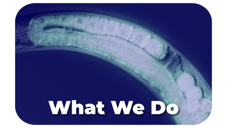
We use the model organism Caenorhabditis elegans to decode the molecular pathways that govern fertility from structure to phenotype.

By defining the molecular parts list and wiring diagram of reproduction in a simple model organism, we are laying the groundwork to better understand the causes of infertility and miscarriage.

We use cutting-edge genome editing technology together with biochemical, biophysical, and sequencing-based methods to define the molecular events required for successful embryo development.

We are a team of dedicated researchers who seek to unlock the mysteries of how living things reproduce.
Asli Ertekin, Sharon T. Noronha, Christable Darko, Francesca Massi, Sean P. Ryder.
RNA, (2025) 31(11):1575-1588
Sexually reproducing organisms make haploid gametes—oocytes and spermatocytes—that combine during fertilization to make an embryo. While both gametes contain similar DNA content, oocytes contain the bulk of the cytoplasm including maternally supplied mRNAs and proteins required prior to zygotic gene activation. RNA-binding proteins are key regulators of these maternal transcripts. In Caenorhabditis elegans, the tandem zinc finger proteins OMA-1 and OMA-2 are required for fertilization. Here, we show that OMA-1 RNA-binding activity requires a short basic region immediately upstream of the canonical tandem zinc finger domain. Mutation of this region in animals produces a phenotype distinct from a genetic null. Oocytes can be fertilized, but fail to form an intact chitin eggshell, frequently fragment in utero, and arrest prior to morphogenesis. Our results identify a critical region outside of the canonical RNA-binding domain required for both RNA-binding activity as well as revealing a new role for OMA-1 during the oocyte-to-embryo transition.
 The mex-3 3' untranslated region is essential for reproduction during temperature stress.
Hannah E. Brown, Haik V. Varderesian, Sara A. Keane, Sean P. Ryder
Development (2025) Aug 11:dev.204740. doi: 10.1242/dev.204740 (online ahead of print)Organisms must sense temperature and modify their physiology to survive environmental stress. Elevated temperature reduces fertility in most sexually reproducing organisms. Maternally supplied mRNAs are required for embryogenesis. They encode proteins that govern early embryonic patterning. RNA-binding proteins (RBPs) are major effectors of maternal mRNA regulation. MEX-3 is a conserved RBP essential for anterior patterning of Caenorhabditis elegans embryos. We previously demonstrated that the mex-3 3´ untranslated region (3´UTR) represses MEX-3 abundance in the germline yet is mostly dispensable for fertility. Here, we show that the 3´UTR is essential during thermal stress. Deletion of the 3´UTR causes a highly penetrant temperature sensitive embryonic lethality phenotype distinct from a mex-3 null. Loss of the 3´UTR decreases MEX-3 abundance specifically in maturing oocytes and early embryos during temperature stress. Dysregulation of mex-3 reprograms the thermal stress response by reducing the expression of hundreds of heat shock genes. We propose that a major function of the mex-3 3´UTR is to buffer MEX-3 expression during fluctuating temperature, ensuring the robustness of oocyte maturation and embryogenesis.
 The endogenous mex-3 3' UTR is required for germline repression and contributes to optimal fecundity in C. elegans
Mennatallah M.Y. Albarqi. and Sean P. Ryder.
PLOS Genet. (2021) Aug 23;17(8):e1009775. doi: 10.1371/journal.pgen.1009775.
x
RNA regulation is essential to successful reproduction. Messenger RNAs delivered from parent to progeny govern early embryonic development. RNA-binding proteins (RBPs) are the key effectors of this process, regulating the translation and stability of parental transcripts to control cell fate specification events prior to zygotic gene activation. The KH-domain RBP MEX-3 is conserved from nematode to human. It was first discovered in Caenorhabditis elegans, where it is essential for anterior cell fate and embryo viability. Here, we show that loss of the endogenous mex-3 3´UTR disrupts its germline expression pattern. An allelic series of 3´UTR deletion variants identify repressing regions of the UTR and demonstrate that repression is not precisely coupled to reproductive success. We also show that several RBPs regulate mex-3 mRNA through its 3´UTR to define its unique germline spatiotemporal expression pattern. Additionally, we find that both poly(A) tail length control and the translation initiation factor IFE-3 contribute to its expression pattern. Together, our results establish the importance of the mex-3 3´UTR to reproductive health and its expression in the germline. Our results suggest that additional mechanisms control MEX-3 function when 3´UTR regulation is compromised.
